Sinking Coffin Bones (Part 1 of 2)
Horseback Magazine
8-10-2013
Pete Ramey
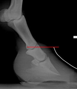
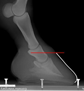
When looking at a lateral radiograph of a horse’s foot, if
the exact location of the top of the hoof wall (hairline) has been marked with a
radiopaque paste or object, there is a measurable distance between the
“elevation” of the top of the hoof capsule and the top of the coffin bone.
Veterinarians and farriers typically refer to this measurement as the CE
(coronet-to-extensor process
distance).
The
markers (taped-on wires) were placed contouring the hoof wall and stopping
precisely at the base of the hairs at the coronet. In figure A, the CE is more
than one inch, with most of the short pastern bone (P2) buried within the hoof
capsule. In figure B, the same hoof six-months-later, the CE is almost within a
normal range (post-treatment). In both photos, the sole thickness is roughly the
same, yet the overall wall length in figure A is dramatically longer than in the
healthier situation of figure B. Photo reprinted from the book Care and
Rehabilitation of the Equine Foot , P. Ramey.
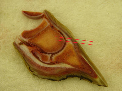
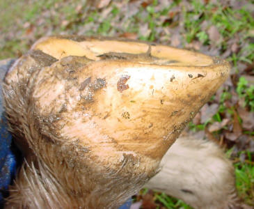
Cross-section of a stillborn foal’s foot. The CE is near zero—the hairline is
almost level with the top of the coffin bone. Photo
reprinted from the book Care and Rehabilitation of the Equine Foot, P.
Ramey.
In newborn horses, and in the healthiest examples of adult
horses, the CE measurement will be near zero. In other words, the top of the
coffin bone will be level with (or within 1/2-inch of) the top of the hoof
capsule (hairline). This “high” (actually normal) bone position allows the
overall hoof length to be very short and compact (usually around 3-3 ½-inches
long at the toe), while still having room for a thick, strong, robust sole
beneath the bone and sensitive tissues.
Over time, many domestic horses literally sink through
their hoof capsule. The CE measurement can grow to an inch or more in horses
that are not extremely lame (though they will not be “right,” either). When the
CE measurement becomes abnormally high (more than 1/2-inch), this means—among
other things—that if the horse is to have an adequately-thick sole, he must also
have a longer-than-normal overall hoof wall length. This, of course, leads to
all sorts of locomotive and performance problems, whether the farrier chooses
to, a) thin the soles to achieve normal
wall lengths, or b) leave adequate sole thickness at the expense of leaving the
extra wall length along with it. Which is correct? Neither. The right choice is
to maintain the CE of your horse between zero and 1/2-inch. Then you can have a
thick sole and a short,
compact hoof capsule.
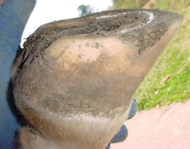
In
figure D (left), the toes (and heels) would be considered too long by any hoof
professional, yet the soles are paper-thin. If this hoof was to be cut
shorter—thus thinning the sole more—it would be severely damaging for the horse.
By, instead, focusing on reversing the coffin bone sinking, the same foot (shown
in figure E, right) now has a thick sole
and a normal heel and toe length (four months duration between photos).
Photo reprinted from the book Care
and Rehabilitation of the Equine Foot, P. Ramey.
Understanding the
Problem
To understand how to reverse or prevent the sinking, you
must first understand how and why it occurs. The coffin bone is shaped like a
miniature hoof, creating the foundation for the front-half of the horse’s foot.
The bone is surrounded by a 1/8-to-1/4-inch “sock” of blood vessels,
nerves, and connective tissue. The hoof wall, around the perimeter, and the sole
underneath forms a tough outer shell—like a boot—about 1/2-inch-thick. In a
natural and healthy situation, the hoof wall and the sole share the load of the
horse’s weight. In this situation, the laminae—the bonds between the hoof wall
and the coffin bone—have little or no shear stress forces applied to them.
However, if the hoof wall is allowed to overgrow well-past the sole, or
if a shoe is lifting the sole off the ground, the forces change dramatically—the
horse’s entire weight is literally hanging from the laminae. These forces set up
two possible scenarios:
1)
If the diet is correctly balanced and the horse
is generally healthy, so that no additional stressors are placed on the
integrity of the laminae, the additional vertical forces applied to the laminae
may allow the horse to slowly sink through the hoof capsule over time. This can
occur without a lot of pain, and can be fairly easy to reverse.
2)
If the horse’s health is compromised, or if an
improper diet is weakening the laminae, the horse may suddenly
fall through the hoof capsule,
essentially until the sole reaches the ground. This may destroy connective
tissue and blood supply to a point that the foot could never be fully healed.
In either case, I feel that the sinking was caused by the
unloading of the horse’s sole to begin with—by placing the laminae in the
solitary support role, without the aid of the rest of the foot (sole, bars,
frogs). This brings us to the
concept of the sole penetration. In
the most extreme laminitis cases, the coffin bone supposedly pierces through the
sole of the horse. Since the sole is skin
that literally grows from the bottom of the coffin bone, I do not understand why
people believe that this “piercing” can occur—the sole is
attached
to the bone, and moves around with it
wherever it goes. If the bone sinks, so must the sole. So instead, I consider
the CE as one issue, and the sole
thickness as a completely separate-but-important issue. In cases that people
believe the bone penetrated the sole; I would, instead, be asking why the sole
is missing. Did the corium abscess
and allow the sole to fall off? Did someone cut it off? Did it wear away? Has it
failed to grow? This may seem like a simple semantics game, but if we ask the
right questions, we are more likely to find the right answers.
Luckily, most cases of coffin bone sinking aren’t quite so
dramatic. Instead, all you will notice over time is that the toes (and/or heels)
seem to be getting longer, or the soles seem to be getting thinner, or both. The
horse is not quite as sound as he used to be—or is not an
easy-mover anymore. If radiographs
verify that the CE is ½- to ¾-inch or more, your horse will benefit from a
conscious effort to reverse the situation. To do this, we basically set up the
opposite of the forces that caused
the situation to begin with. We try to reduce the load on the walls, while
increasing the load on the rest of the foot. This means frequent trimming of the
walls, conservation of the sole and frog tissue, and using hoof boots with
padded insoles to compensate for the reduced support that would normally be
provided by longer hoof walls.
Since the laminae are weakened by sugar overload and/or
mineral imbalances, we also carefully balance the diet. This gives the best
chance of success by helping the wall connection
be the best it can be.
Next Month: How to
Reverse Coffin Bone Sinking (part 2 of 2)